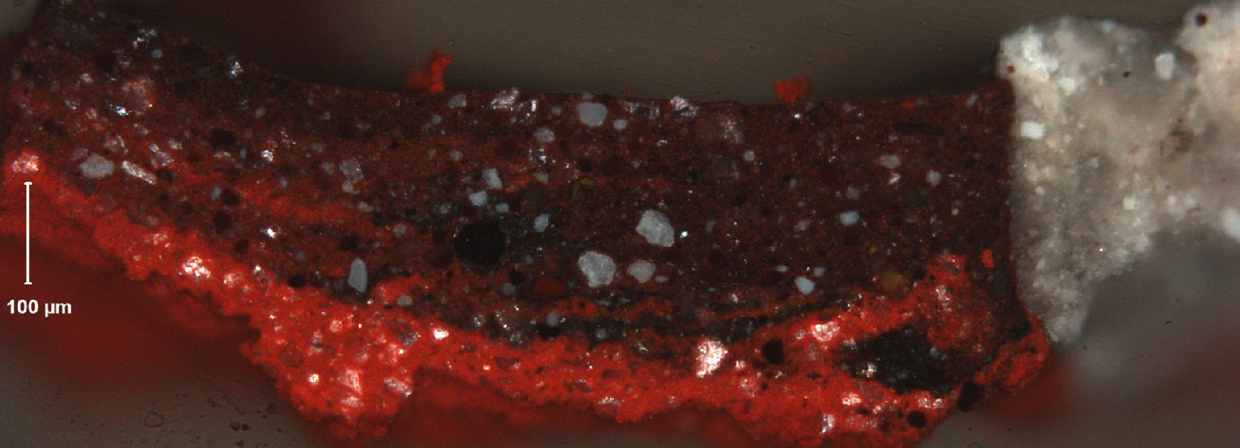
By Pam Skiles, Associate Conservator of Paintings at the Denver Art Museum
One of the ways that conservators can learn more about paintings is through cross sections. These are very small samples, no bigger than the head of a pin, and they can provide information to identify pigments and media. The samples are taken from the painting and mounted onto slides to be examined under a microscope. Using a microscope and visible light, the “stratigraphy” (layers of paint) of the paint layers is revealed and these layers can be examined in great detail. By viewing the samples using other energies, like ultraviolet radiation, conservators can see details not visible in normal light. Ultraviolet radiation is used to characterize the media and pigments, which helps guide treatment decisions.
Additionally, these samples can be analyzed with a scanning electron microscope (SEM) to get an extremely detailed image. Instead of using light to illuminate the sample, the SEM bombards the sample with electrons, which creates the image. From these images, elemental analysis can be carried out and helps determine which paints the artist used.


